Categories


- 0086-512-58697986
- info@bestrantech.com
- bestrantech
- 0086-13862268429
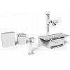
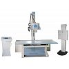
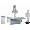
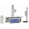
High Frequency Digital Radiography System

BT-XR11 High Frequency Digital Radiography System
I. Application:
This machine is applied to take radiography on every part of human body, such as head, limbs, chest, limbus and abdomen and etc.
II. Specification
|
High- frequency X-ray machine |
Output power |
50kW |
|
|
Main inverter frequency |
260kHz |
||
|
X-ray tube |
Dual-focus X-ray tube |
Small focus:0.6 Large focus:1.2 |
|
|
Output power |
22kW/50 kW |
||
|
Anode Capacity |
150kHU |
||
|
Cooling |
180W |
||
|
Anode Angle |
12° |
||
|
3200rpm |
|||
|
Tube Current |
10mA- 650mA |
||
|
Tube voltage |
40-150Kv |
||
|
mAs |
1-1000mAs |
||
|
Exposure Time |
0.001-6.3s |
||
|
AEC |
Optional |
||
|
Digital Image System |
Digital Detector |
Field of view |
17”*17” |
|
Pixel |
3K*3K |
||
|
Ultimate spatial resolution |
3.7LP/mm |
||
|
Pixel size |
143um |
||
|
Output grayscale |
14bit |
||
|
Imaging time |
9s |
||
|
DQE |
70% |
||
|
Image Workstation |
Acquisition module |
Inside enhancement module |
|
|
Image information management |
Dicom image transmission Dicom film printing Dicom image storage (hard disk, compact disk) |
||
|
Mechanical structure and performance |
U-arm |
Vertical movement range |
≥1250 mm(motorized control) |
|
Focus-screen movement range |
1000mm-1800mm(motorized control) |
||
|
Rotation range |
-40°-+130°(motorized control) |
||
|
Detector rotation |
-45°-+45° |
||
|
Tube rotation |
180° |
||
|
Photography table (Optional) |
Table size |
2000mm*650mm |
|
|
Table height |
≤740mm |
||
|
Transverse movement |
200mm(electromagnetic lock) |
||
|
Longitudinal movement |
100mm(electromagnetic lock) |
||
|
Power supply |
380V 50/60Hz |
||
III. Product Details
1. Type of generator and X-ray tube:
l Advanced 260kHz high frequency high voltage type generator, realize 1ms instant exposure, high performance.
l Three exposure method free change: KV, mAs two adjustment, KV, mA, s three adjustment and AEC function(optional), to satisfy different habit of different doctors.
l Rotate double anode 0.6/1.2, with high heat capacity of 150KHU
l Digital micro-processed closed loop control and malfunction alarming system to reduce the dose of X-ray protect the patients and doctors very well.
l LCD touch screen, beautiful appearance and convenience to operate.
2. Flat Panel Detector
l Apply with A-Si (Amorphous silicon) Toshiba imported Flat panel detector, which could give perfect digital images directly.
l 3K×3K Acquisition matrix, 143um pixels size, and 3.7Lp/mm ultimate spatial resolution, with DQE values ≥ 70%
l Shortest imaging time ≤ 9s
l 17〞×17〞Large acquisition area and with the non-center processing technology, no matter the center and border, the quality of the image is the same.
l The detector could be rotated ±45 degree along the axis direction, to satisfy the different photograph requirement of every body parts, such as Ankle joint, lateral spine
l The detector has the self-protection function. It can stop to move when it detect the distance in front of the barrier.
3. Digital Working Station:
|
|
Case registration: Auto registration, be equipped with Dicom Worklist SCU. To simplify the input process for doctors, greatly reduce the amount of labour and greatly improve the working efficiency
l Image Acquisition: Automatic window adjustment, Automatic cropping, Automatic transmit.
l Image Processing: Tissue equilibrium, W/L adjustment, Gamma correction, interest district, reversed phase, noise reduction, smooth, sharpen, pseudo color, edge extraction, shadow compensation, filter nuclear, single window, dual-window, four windows, movement, right rotated 90°, left rotated 90°, level mirror image, vertical mirror image, magnifying glass, image zooming, reset, layer information, label character, drawing label, length measurement, angle measurement, rectangular length, rectangular area, elliptic length, elliptic area.
l Dicom Image Transmit, Dicom Image Storage, Dicom Image viewing, Dicom Image printing.
l Convenient to connected to the PACS system
4. Operation System:
l Be equipped with 24〞imported LCD high resolution monitor screen, the delicate and richness degree of image is far higher than the normal medical monitor. International advanced level.
l Brightness and Contrast are higher than 1000NIT, far higher than the normal LCD screen of 400 NIT.
l These features can make the doctor diagnose more accurate and smooth.
l Be equipped with the microphone and remote exposure control. The doctor can control outside the operating room.
l Be equipped with various set of infrared facilities to protect the machine from the mis-operation of the doctors.
l Optional PLXF153 operating room. Battery power supplied, Infrared unlock
l Optional SONY, CODONICS film printer.
5. Mechanical Movement:
l The self-designed and manufactured electric U-arm mainframe can move up and down, and rotate in a wide range, which can satisfy the requirements of multi-site photography.
l Adopting original Italian geared motor, the features are reliable performance, lower noise, longer service life.
IV. Standard Configurationt
|
No. |
Item |
Quantity |
|
1 |
Newly designed U-arm frame |
1 |
|
2 |
High frequency and high voltage X-ray generator |
1 |
|
3 |
X-ray tube |
1 |
|
4 |
17’’*17” digital detector |
1 |
|
5 |
Image processing workstation |
1 |
|
6 |
24” High-definition LCD display |
1 |
|
7 |
Human graphical touch screen |
1 |
|
8 |
High frequency inverted power supply electronic cabinet |
1 |
|
9 |
Symmetry adjustable collimator with light |
1 |
V. Options
1. BT-XA04 Intelligent All-Directions Mobile Table
(2000×650×740mm, Longitudinal movement: 200mm, Horizontal movement: 100mm)